Anthropological data to the Árpádian Age – Examination of Jászberény Szent Pál-halom (11–13th century)
Abstract
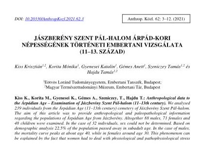
We analysed 239 individuals from the Árpádian Age (11–13th century) cemetery of Jászberény Szent Pál-halom. The aim of this article was to provide anthropological and paleopathological information regarding the populations of Árpádian Age from Jászberény. Altogether 88 males, 71 females and 48 children were examined. In the case of 32 individuals, sex could not be determined. Based on demographic analysis 22.5% of the population passed away in subadult age. In the case of males, the mortality curve peaks at about age 40, while in females around age 30. This phenomenon can be explained by the fact that women had to deal with physiological and pathophysiological stress due to pregnancy and its complications. The average height was 167.11 cm for men and 158.07 cm for women. Craniometric analysis revealed some differences between the two sexes, e.g. females were mainly mesokran, while males had hyperdolichokran, dolichokran, mesokran and brachykran skull as well. Porotic hyperostosis was identified most frequently on the orbital roof. Signs of premortem and postmortem traumas were also identified. Interpersonal violence is indubitable as premortem cut marks were observed in more cases. The premortem traumas were mostly related to lifestyle, possible accidents and cultural intentions. Linear enamel hypoplasia was most common on the lower first and second incisors, furthermore on the lower canines. The prevalence was much higher in males compared to females.
References
Alekszejev, V.P., Debec, G.F. (1964): Kraniometrija. Izd. Nauka, Moszkva. pp. 128
Aufderheide, A.C., Rodríguez-Martín, C. (1998): The Cambridge encyclopedia of human paleopathology. Cambridge University Press, Cambridge. pp. 478.
Bernert, Zs. (2005a): Paleoantropológiai programcsomag. Folia Anthropologica, 3: 71–74.
Bernert Zs. (2005b): Érvek a testmagasság osztálykategóriák korrekciójának szükségességéről a Kárpát–medencei történeti embertanban. In: Korsós, Z. (Szerk.): V. Kárpát–medencei Biológiai Szimpózium. Előadások összefoglalói. Budapest. pp 33–43.
Bernert, Zs., Évinger, S., Hajdu, T. (2007): New data on the biological age estimation of children using bone measurements based on historical populations from the Carpathian Basin. Annales historico-naturales Musei nationalis hungarici, 99: 199–206.
Bernert, Zs., Évinger, S., Hajdu, T. (2008): Adatok a gyermekek életkorbecsléséhez a Kárpát-medencei történeti népességek gyermekhalottainak csontméretei alapján. Anthropologiai Közlemények, 49: 43–50.
Brooks, S., Suchey, J.M. (1990): Skeletal age determination based on the os pubis: a comparison of the Acsádi-Nemeskéri and Suchey-Brooks methods. Human Evolution, 5: 227–238. DOI: https://doi.org/10.1007/BF02437238
Coale, A.J., Demény, P. (1966): Regional modell life tables and stable populations. Princeton University Press, Princeton, USA. pp. 871.
Csalog, Zs. (1964): Jászberény-Szentpálhalom. In: Sz. Burger, A. (Szerk.) Az 1963. Év Régészeti Kutatásai. Régészeti Füzetek. 17. Magyar Nemzeti Múzeum - Történeti Múzeum, Budapest.
Éry, K. (1982): Újabb összehasonlító statisztikai vizsgálatok a Kárpát-medence 6–12. századi népességeinek embertanához. Veszprém megyei Múzeumok Közleményei, 16: 35–86.
Éry K. (1998): Length of limb bones and stature in ancient populations in the Carpathian basin. Humanbiologia Budapestiensis, 26: pp. 96.
Éry, K., Kralovánszky, A., Nemeskéri, J. (1963): Történeti népességek rekonstrukciójának reprezentáció. Anthropologiai Közlemények, 7: 41–90.
Fazekas, G., Kósa, F. (1978): Forensic fetal osteology. Akadémiai Kiadó, Budapest. pp. 414.
Ferembach, D., Schwidetzky, I., Stloukal, M. (1979): Empfehlungen für die Alters- und Geschlechtsdiagnose am Skelett. Homo, 30: 1–32.
Iscan, M.Y., Loth, S.R., Wright, R.K. (1984): Age estimation from the rib by phase analysis: white males. Journal of Forensic Sciences, 29: 1094–1104. DOI: https://doi.org/10.1520/JFS11776J
Iscan, M.Y., Loth, S.R., Wright, R.K. (1985): Age estimation from the rib by phase analysis: white females. Journal of Forensic Sciences, 30: 853–863. DOI: https://doi.org/10.1520/JFS11018J
Józsa, L., Pap, I. (1991): Vashiányos anaemia a honfoglalás és az Árpádok korában. Orvosi Hetilap, 28: 1544–1545.
Kaposvári, Gy. (1960): Jászberény-Szentpál halom. In: Az 1959. Év Régészeti Kutatásai. Régészeti Füzetek 13. Magyar Nemzeti Múzeum – Történeti Múzeum, Budapest.
Loth, S.R, Henneberg, M. (1996): Mandibular ramus flexure: a new morphologic indicator of sexual dimorphism in the human skeleton. American Journal of Physical Anthropology, 99: 473–485. DOI: https://doi.org/10.1002/(SICI)1096-8644(199603)99:3<473::AID-AJPA8>3.0.CO;2-X
Lovell, N. (2008): Analysis and interpretation of skeletal trauma. In: Katzenberg, M.A., Grauger, A.L. (Ed.) Biological anthropology of the human skeleton. John Wiley & Sons, New York. pp. 341–386.
Mann, R.W., Hunt, D.R. (2005): Photographic Regional Atlas of Bone Disease – A Guide to Pathologic and Normal Variation in the Human Skeleton. Charles C. Thomas Publisher, Springfield. pp. 432.
Mann, R.W., Jantz, R.L., Bass, W.M., Willey, P.S. (1991): Maxillary suture obliteration: a visual method for estimating skeletal age. Journal of Forensic Sciences, 36: 781–791. DOI: https://doi.org/10.1520/JFS13088J
Marcsik, A. (1975): Presumed etiology of a bone change. Anthropologiai Közlemények, 19: 47–53.
Martin, R., Saller, K. (1957): Lehrbuch der Anthropologie. Bd. 1. Gustav Fischer Verlag, Stuttgart.
Meindl, R.S., Lovejoy, C.O. (1985): Ectocranial suture closure: a revised method for the determination of skeletal age at death. American Journal of Physical Anthropology, 68: 57–66. DOI: https://doi.org/10.1002/ajpa.1330680106
Nikita, E. (2017): Ostearcheology. A Guide to the Macroscopic Study of Human Skeletal Remains. Academic Press, London. pp. 462.
Ortner, D.J. (2003): Identification of a pathological conditions in human skeletal remains. Academic Press, San Diego. pp. 605.
Pilloud, M.A., Canzonieri C. (2014): The occurrence and possible aetiology of spondylolysis in a pre-contact California population. International Journal of Osteoarchaeology, 24: 602–613. DOI: https://doi.org/10.1002/oa.2245
Rogers, T.L. (1999): A visual method of determining the sex of skeletal remains using the distal humerus. Journal of Forensic Sciences, 44: 57–60. DOI: https://doi.org/10.1520/JFS14411J
Schinz, H.R., Case, J.T. (1952): Roentgen-diagnostics. Grune & Stratton, New York.
Schour, I., Massler, M. (1941): The development of the human dentition. Journal of the American Dental Association, 28: 1153–1160.
Sjøvold, T. (1990): Estimation of stature from long bones utilizing the line of organic correlation. Journal of Human Evolution, 5: 431–444. DOI: https://doi.org/10.1007/BF02435593
Stloukal, M., Hanáková, H. (1978): Die Lange der Langsknochen altslawischer Bevölkerungen unter besonderer Berücksichtigung von Wachstumsfragen. Homo, 29: 53–69.
Ubelaker, D.H. (1989): Human skeletal remains: excavation, analysis, interpretation. 2nd ed. Taraxacum, Washington. pp. 172
Waldron, T. (2008): Paleopathology (Cambridge Manuals in Archeology). Cambridge University Press, Cambridge. pp. 298.
Wentz, R., De Grummond, N. (2009): Life on horseback: palaeopathology of two Scythian skeletons from Alexandropol. International Journal of Osteoarchaeology, 19: 107–115. DOI: https://doi.org/10.1002/oa.964


